

Title: IHC staining of an EAE (model of multiple sclerosis) lesion in a murine spinal cord
Votes: 15
Views: 580
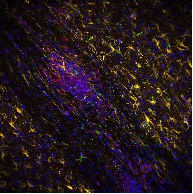
Description: This image is of an EAE lesion in a murine spinal cord at a chronic, inflammatory timepoint (d40) in disease. Astrocytes (yellow) are seen to extend their processes and envelop CD45+ cells (green); thereby suggesting interaction between the two at the rim of the lesion. This is critical as little is known about astrocyte interactions with infiltrating immune cells in the context of Multiple Sclerosis. IHC staining for CD45 (green), GFAP (yellow), and C3 (red) in the spinal cord of a EAE treated mouse at a chronic (d40) inflammatory timepoint. Image is of a EAE lesion characteristic of lesions in MS tissue. CD45 is a general immune cell marker, GFAP is a general astrocyte marker, and C3 is an inflammatory protein. Image taken on SP8 confocal microscope at 25x