

Title: Microglial turf war in the auditory midbrain
Votes: 6
Views: 561
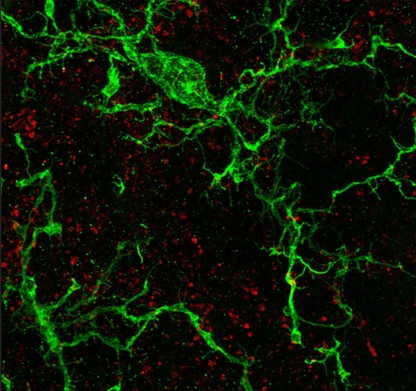
Description: Confocal micrograph showing a maximum intensity projection of iba1+ microglia (green) and synaptophysin+ putative pre-synapses (red). Imaged on a Leica Stellaris 5 confocal with Lightning super-resolution at Manchester Metropolitan University in October 2022. Microglial cells in the healthy brain tile the parenchyma in non-overlapping territories. One role for microglia involves the active movement of ramified processes which interact with synapses. We are investigating the fundamental nature of these interactions in the inferior colliculi, the principal midbrain nuclei in the mammalian auditory pathway. This image shows two ramified microglia with non-overlapping territories that are surrounded by punctate pre-synapses. NB - due to the maximum intensity projection of the z-stack shown, some pixels appear to colocalise both labels (yellow). However, in the individual slices of the z-stack, these can be seen to be points where microglia abut but do not colocalise with synapses, demonstrating the importance of super-resolution and single plane analyses of z-stacks in these experiments. Contact l dot orton at mmu dot ac dot uk.