

Title: Tentacles of the Neuron
Votes: 702
Views: 3417
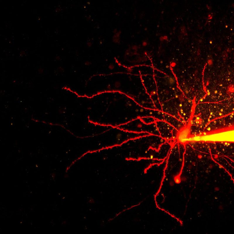
Description: This image shows a hippocampal CA1 neuron which is whole-cell patched using a pipette containing the internal solution along with a calcium insensitive dye (red, Alexa 594) and a calcium sensitive dye (green, Fluo 4F). You can clearly see the dendritic tree and the spines on the dendrites. Also seen is an axonal bleb (red, round structure at the end of the axon) that is formed following slicing procedures.