

Title: GFAP-ulous
Votes: 30
Views: 320
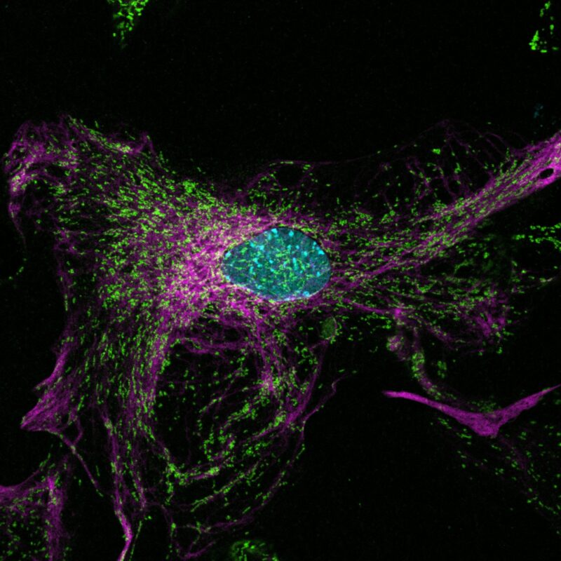
Description: This image was captured using an Olympus FLUOVIEW FV3000 laser confocal microscope with five laser lines (405, 488, 561, 594, 640). Primary astrocyte cultures from P1-P4 mice were fixed with PFA and stained using immunocytochemistry. Astrocytes were labeled with GFAP (Glial Fibrillary Acidic Protein, 1:1000), a cytoskeletal marker for astrocytes, and TOM20 (Translocase of the Outer Membrane 20, 1:200), a mitochondrial membrane protein. Secondary staining was performed with Alexa Fluor 647 (GFAP) and Alexa Fluor 546 (TOM20). DAPI mounting media was used to stain nuclei. The sample was imaged at 30x with 2x zoom (2048x2048 resolution). Post-processing involved contrast and brightness adjustments in FIJI. The final image reveals the intricate mitochondrial architecture (green) within astrocytic processes (magenta).