
Title: Glial Components of the Blood-Brain Barrier in Human FCD-Associated Epilepsy
Votes: 44
Views: 467
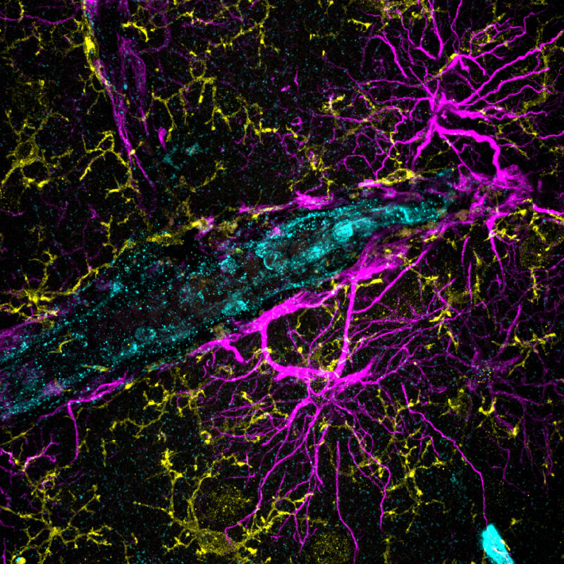
Description: This image captures the neurovascular unit within a block of the right temporal cortex, specifically from the ictal zone of a human patient diagnosed with focal cortical dysplasia type II (FCD2). The tissue was removed during surgical resection for drug-resistant epilepsy. Imaging was performed using a Nikon C2 confocal laser scanning microscope with a 60× oil-immersion objective, using z-stack acquisition to reconstruct the 3D organization of cells and blood vessels. In the image, the blood vessels are shown in turquoise, microglia in yellow, and astrocytes in magenta, with their perivascular endfeet "touching" the vessel's surface. This image highlights essential glial elements of the neurovascular unit within an epileptogenic cortical zone, where atypical glial–vascular interactions may contribute to blood-brain barrier dysfunction, chronic inflammation, and neuronal hyperexcitability characteristic of FCD-associated epilepsy.