
Title: EV Kaleidoscope: Unveiling Colourful Secrets of the Brain
Votes: 3
Views: 161
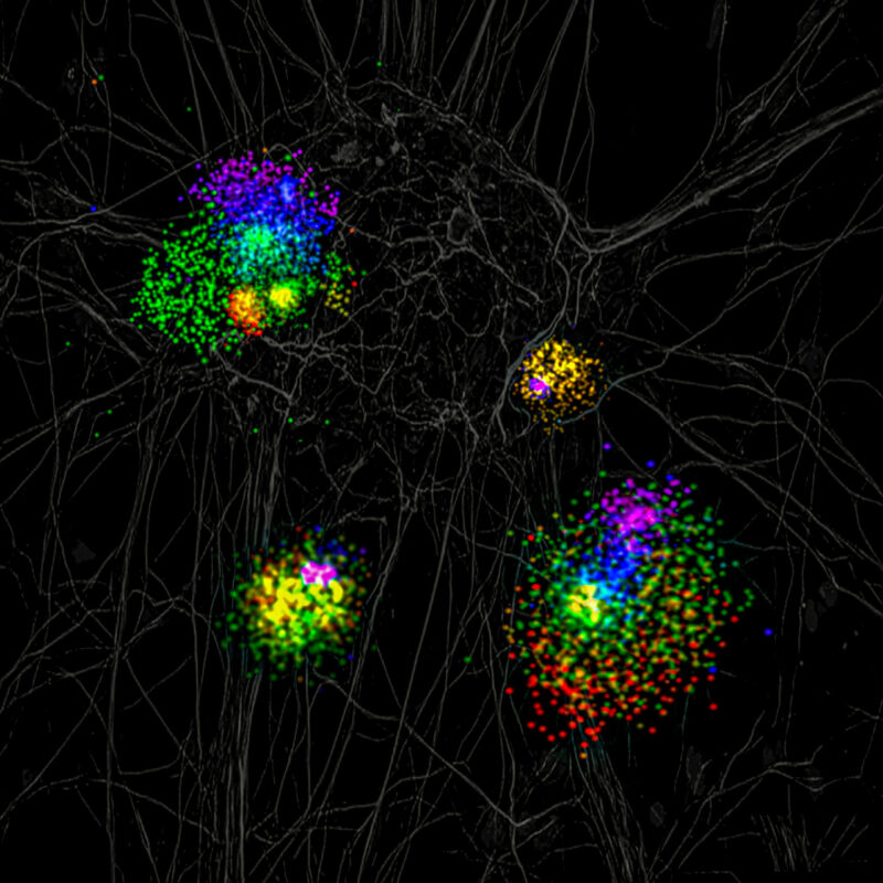
Description: This image was created by combining super-resolution dSTORM images of four extracellular vesicles (EVs), each captured at the highest magnification from different fields of view. Due to their minuscule size (50–200 nm), zooming into a single field would result in a loss of resolution. To ensure clarity, individual EVs were combined into a single field of view. Basic color and contrast adjustments were made to enhance key features, and the EV images were overlaid onto a grayscale background resembling neurons to emphasize their connection to brain function and disease, all while maintaining the scientific integrity of the image. Extracellular Vesicles [EVs] are produced and released by all cells in our body. These minuscule cellular messengers play crucial roles in intercellular communication and are essential for various biological functions, including disease signalling. EVs contain cell- and, most importantly, cell-state specific cargo, which is the reason why they are considered as potential source of disease biomarkers. In this image, I delve into the intricate world of EVs, isolated from frozen post-mortem brain tissue of a patient afflicted with dementia. While analysis of EV cargo often requires highly sensitive analytical methods like single-molecule array (Simoa), electrochemiluminescence EIA, luminex assay etc., until recently, visualizing EVs at nanoscale was an insurmountable challenge, with transmission electron microscopy as our sole ally. This cutting-edge super-resolution microscopy image unveiled a mesmerizing canvas of EVs, appearing as molecular fireworks. The nano-vesicles were labelled with antibodies to common EV surface markers CD9 and CD81, and a neurodegenerative disease-causing protein alpha Synuclein, each tagged with three different fluorophores. While the common EV markers appear to be distributed throughout the surface, the alpha Synuclein protein appeared as clusters inside EVs. This frameindex dSTORM image stands as a testament to the capabilities of super-resolution microscopy, illuminating the uncharted territory within a single vesicle showing individual markers in different colors. Depending on the state (monomer vs aggregates) and location (inside vs on the EV membrane) of this protein, insights into its potential role as a therapeutic target or disease biomarker can be refined. Therefore, this technique carries vast potential in discerning disease biomarkers residing within individual EVs, ushering in a new era of diagnostic precision.