

Title: pig's motor cortex
Votes: 345
Views: 2313
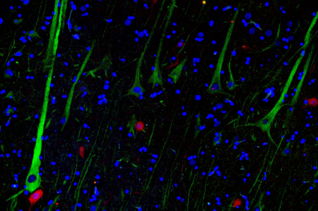
Description: Immunofuorescence colocalization was performed on serial 5-μm sections of pig's motor cortex using a rabbit polyclonal anti-MAP2 antibody (ab32454, 1:500) as pyramidal neurons marker (green) and a mouse monoclonal anti-Parvalbumin (PV) (Sigma, P3088, clone Parv-19, 1:1000) as interneurons marker (red). Epitope retrieval was carried out at 120 °C in a pressure cooker for 5 min with a Tris/EDTA bufer, pH 9.0. Sections were blocked for 1 h with 5% normal horse serum (PK-7200, Vector Labs) in PBS and then incubated overnight at 4 °C in a solution of anti-MAP2 and anti-PV in PBS containing 2% normal horse serum and 0.05% Triton X-100. Sections were then rinsed in PBS (3 × 10 min), incubated for 1 h at room temperature in a solution of DyLight 488 anti-rabbit IgG (5 μg/mL, DI-1088; Vector Labs., Burlingame, CA, USA) and DyLight 594 anti-mouse IgG (5 μg/mL, DI-2594; Vector Labs., Burlingame, CA, USA). Finally, sections were washed with PBS and cover slipped with Vectashield medium containing 4′,6-diamidino 2-phenylindole (DAPI) (H-1500, Vector Labs). Microphotographs were collected under a Nikon Ni-E light microscope (Nikon Instruments, Spa Calenzano, Florence, Italy), fully equipped for fluorescence acquisition, connected to a personal computer via Nikon digital image processing software (Digital Sight DS-U1, NIS-Elements BR 4.51.00 software)