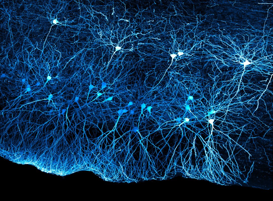


Title: Lateral Entorhinal Cortex
Votes: 18
Views: 501
Description: Confocal image of principal neurons in LII and LIII lateral entorhinal cortex of a mouse brain. Sparse labeling was performed through an injection of AAVretro-CAG-GFP into hippocampus. A thick section (400um) was prepared and cleared using methyl salicylate, and the image was acquired with a 20x objective as a Z-stack (approx. 190um). The image is shown as a maximum intensity projection, using a 'cyan-to-white'; intensity LUT.